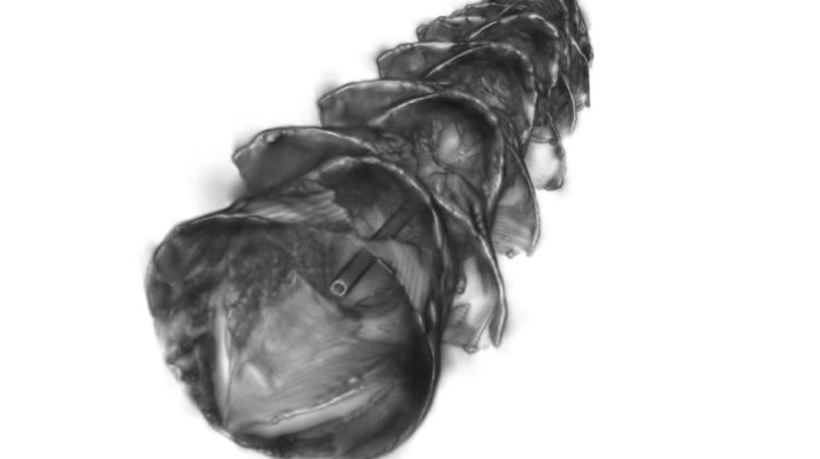
A team of engineers has designed a new type of stent that could be used to deliver drugs to the gastrointestinal tract, respiratory tract and other tubular organs. The engineers took inspiration from kirigami, the Japanese art of folding and cutting paper to create three-dimensional structures.
Thesoft, stretchy tubular stents are made of silicone-based rubber that has been coated in a smooth layer of plastic that has itself been etched with small ‘needles’ that pop up when they’re stretched. The needles penetrate the surrounding tissue and deliver a payload of drug-containing microparticles. Once the stent has been removed, the drugs continue to be released over an extended period.
According to one of the study’s authors, Giovanni Traverso, an assistant professor of mechanical engineering at Massachusetts Institute of Technology and a trained gastroenterologist, this kind of localised drug delivery could make it easier to treat inflammatory diseases affecting the gastrointestinal tract, such as inflammatory bowel disease or eosinophilic esophagitis, by delivering drugs directlyto the affected tissues.
‘This technology could be applied in essentially any tubular organ,’ Traverso said. ‘Having the ability to deliver drugs locally, on an infrequent basis, really maximises the likelihood of helping to resolve patients’ conditions and could be transformative in how we think about patient care by enabling local, prolonged drug delivery following a single treatment.’
Inserting stents into the gastrointestinal tract can be difficult because digested food is continuously moving through the tract. The MIT team sought to get around this by creating a stent that would be inserted temporarily, lodge firmly into the tissue to deliver its payload and then be easily removed.
‘The novelty of our approach is that we used tools and concepts from mechanics, combined with bioinspiration from scaly-skinned animals, to develop a new class of drug-releasing systems with the capacity to deposit drug depots directly into luminal walls of tubular organs for extended release,’ said the study’s lead author, Sahab Babaee, an MIT research scientist. ‘The kirigami stents were engineered to provide a reversible shape transformation: from flat, to 3D, buckled-out needles for tissue engagement, and then to the original flat shape for easy and safe removal.’
Researchers in Traverso’s lab have previously used kirigami to design a non-slip coating for shoe soles.
The researchers created kirigami needles with a range of different sizes and shapes. By varying these features, as well as the thickness of the plastic sheet, they found that they could control how deeply the needles penetrate the tissue. ‘The advantage of our system is that it can be applied to various length scales to be matched with the size of the target tubular compartments of the gastrointestinal tract or any tubular organs,’ Babaee said.
The researchers tested the stents by endoscopically inserting them into the oesophagus of pigs. Once in place, the stents were inflated using a balloon inside the stent, which caused the needles to pop up and penetrate roughly half a millimetre into the surrounding tissue. The needles were coated with microparticles containing a steroid used to treat inflammatory bowel disease.
Deflating the balloon caused the needles to flatten out, enabling the researchers to remove the stent endoscopically. The entire process took only a couple of minutes. The microparticles gradually released budesonide for about a week.
The researchers are now experimenting with the delivery of other types of drugs and working on scaling up the manufacturing process, with the goal of eventually testing the stents in human patients.
The study has been published in the journal Nature Materials.


