
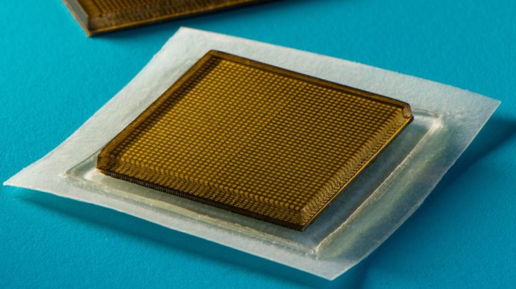
A team of MIT engineers has designed a stamp-sized adhesive patch that can produce continuous ultrasound imaging of internal organs for 48 hours.
Ultrasound imaging currently requires bulky, specialised equipment available only in hospitals and doctor’s offices. In contrast, the new design could potentially make the technology as wearable as a Band-Aid – and as simple to buy.
In the current design, the stickers must be connected to instruments that translate the reflected sound waves into images. According to the researchers, the stickers could have immediate applications even in their current form, such as for continuously imaging internal organs without the need for a technician to hold a probe in place for extended periods.
The team is currently attempting to make the devices work wirelessly. If they are successful, the ultrasound stickers could be made into wearable imaging products that patients could take home from a doctor’s office or even buy at a pharmacy.
Advertisement

‘We envision a few patches adhered to different locations on the body, and the patches would communicate with your mobile phone, where AI algorithms would analyse the images on demand,’ said the study’s senior author, Xuanhe Zhao, professor of mechanical engineering and civil and environmental engineering at MIT. ‘We believe we’ve opened a new era of wearable imaging. With a few patches on your body, you could see your internal organs.’
Currently, ultrasound imaging involves a technician applying a liquid gel to a patient’s skin to transmit ultrasound waves. A probe, or transducer, is then pressed against the gel, sending sound waves into the body that echo off internal structures and return to the probe, where they are translated into visual images.
For patients who require long periods of imaging, some hospitals offer probes affixed to robotic arms that can hold a transducer in place without tiring, but the ultrasound gel flows away and dries out over time, interrupting the imaging.
In recent years, scientists have explored designs for stretchable ultrasound probes that would provide portable, low-profile imaging of internal organs. These devices have a flexible array of tiny ultrasound transducers, enabling them to stretch and conform to a patient’s body. But these experimental designs have produced low-resolution images, in part due to their stretchiness – as they move with the body, the transducers shift location relative to each other, distorting the resulting image.
‘A wearable ultrasound imaging tool would have huge potential in the future of clinical diagnosis. However, the resolution and imaging duration of existing ultrasound patches is relatively low, and they cannot image deep organs,’ said Chonghe Wang, an MIT graduate student.
Advertisement
By pairing a stretchy adhesive layer with a rigid array of transducers, the MIT team’s new ultrasound sticker produces higher-resolution images over a longer duration. ‘This combination enables the device to conform to the skin while maintaining the relative location of transducers to generate clearer and more precise images.’ Wang said.
The device’s adhesive layer is made from two thin layers of elastomer that encapsulate a middle layer of solid hydrogel, a mostly water-based material that easily transmits sound waves. Unlike traditional ultrasound gels, the MIT team’s hydrogel is elastic and stretchy.
‘The elastomer prevents dehydration of hydrogel,’ said Xiaoyu Chen, an MIT postdoc. ‘Only when hydrogel is highly hydrated can acoustic waves penetrate effectively and give high-resolution imaging of internal organs.’
The bottom elastomer layer is designed to stick to skin, while the top layer adheres to a rigid array of transducers that the team also designed and fabricated. The sticker has an area of about two square centimetres and is three millimetres thick.
The researchers ran the ultrasound sticker through a battery of tests with healthy volunteers. The stickers stayed attached, and produced clear images of underlying structures, for up to 48 hours, during which time, the volunteers performed a variety of activities in the lab, from sitting and standing to jogging and lifting weights.
From the stickers’ images, the team was able to observe the changing diameter of major blood vessels when seated versus standing. The stickers also captured details of deeper organs, such as how the heart changes shape as it exerts during exercise. The researchers were also able to watch the stomach distend then shrink back as volunteers drank juice and then passed it out of their system. As some volunteers lifted weights, the team could detect bright patterns in underlying muscles, signalling temporary microdamage.
‘With imaging, we might be able to capture the moment in a workout before overuse and stop before muscles become sore,’ said Chen. ‘We do not know when that moment might be yet, but now we can provide imaging data that experts can interpret.’
The research has been published in Science.